What is duplex ultrasound?
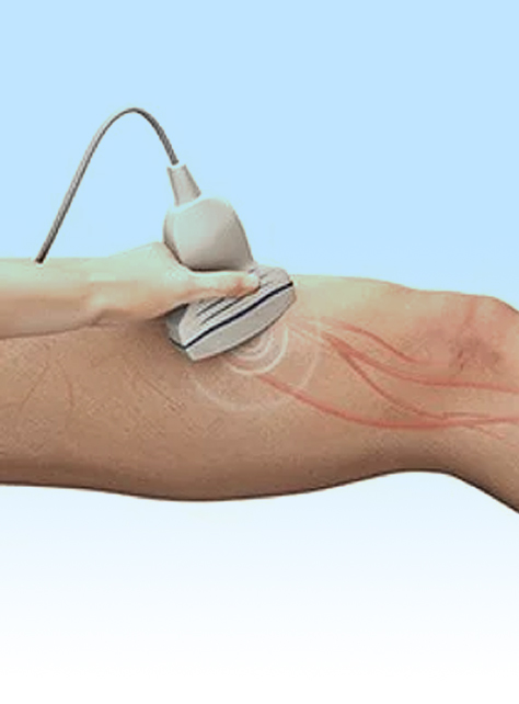
Duplex ultrasound is an imaging technique that is ideal for looking at arteries and veins and how the blood flows through them. Ultrasound uses a transducer to emit sound waves, and based on how those waves return and are measured by the same transducer, the machine produces an image that can be interpreted. There are many different ultrasound modes, and “duplex ultrasound” combines the basic mode and Doppler ultrasound. The combination of these is what gives information of both the structure of the vessels (through the basic mode) and the flow of blood (through Doppler ultrasound). It is fast, low-cost, and does not use any radiation to produce the informative images.
How is duplex ultrasound performed?
To perform a duplex ultrasound, the ultrasound transducer needs to be in direct contact with your skin, so you may be asked to put on a gown to keep your legs exposed. Then, the imaging specialist or technician will likely have you lie down in a position that is comfortable for you. Once you are in position, the specialist or technician will follow your veins or arteries with the ultrasound transducer while saving images and videos. A cool gel will be used to help ensure the transducer obtains good images, and this is usually the only discomfort you should expect. The whole process usually takes between 15-30 minutes.
Duplex Ultrasound
What is duplex ultrasound used for?
Duplex ultrasound is a great tool in the evaluation of chronic venous disease or peripheral artery disease. Because it looks at the structure and blood flow in your vessels, it can tell your doctor which vessels are diseased and to what degree the disease has progressed. This information is valuable in guiding the treatment options that are ideal for your specific case.


